Digestion is the enzymatic decomposition of large molecules of organic components (proteins, fats, carbohydrates) into the low-molecular substances, absorbed by the body and cell membranes. Proper nutrition, that is digestion and absorption of food, requires proper coordination of complex motor activity and secretion of dozens of types of food ingredients degradative enzymes. Digestive system begins with the mouth, then through the throat and esophagus the food reaches stomach and passes into the small intestine. Undigested remains are moved through the intestine to the anus. The length of this road is about 8-9 meters. Sections of the gastrointestinal tract have some common features. They consist of three layers: from the center it is a mucous membrane, the muscle membrane and serous membrane (outer). The differences relate primarily to the construction of epithelium covering the mucosa. Where foods are not absorbed, but only transported, there is a squamous epithelium protective cover. In the stomach and intestines there is a monolayer cylindrical epithelium, covered with an additional seam in the intestine striatum. This type of epithelium is involved both in the absorption of foods and secretory function (digestive juices). Digestive juices are secreted by many digestive glands. They are made up out of the water including enzymes, electrolytes and various organic and inorganic components.
|
|
|
Mouth
In the mouth it comes to the first modification of food: chewing, crushing, mixing with saliva and its moisturizing and partial digestion. Teeth, tongue and salivary glands are involved in that process. Between lips and teeth there is so called vestibule of the mouth, just behind the teeth there is the right mouth. Recall that an adult has 32 permanent teeth (before there is 20 milk teeth). The largest of the salivary glands is located directly to the front of auricle, Parotid gland. Next in size is Submandibular gland, located at the bottom of the mouth. The last large-secreting gland is Sublingual gland.
Esophagus
The next part in the way of food is the esophagus. Is like a continuation of the throat and therefore can also be described as a musculo-membranous cord. The esophagus is a flexible tube with smooth walls, made of muscle and mucous membrane padded from the interior. It is located on the back wall of the chest and after passing through the diaphragm enters the stomach. It connects the throat with the stomach. In the esophagus there is a pressure lower than atmospheric. Absorption of food does not occur there. After swallowing a bite of food is moved into the stomach through the synchronous movement of the muscles of the esophagus called a peristaltic wave. Therefore, transport activity of the esophagus is undergoing even if the throat is located lower than the stomach e.g. while hanging upside down.
Stomach
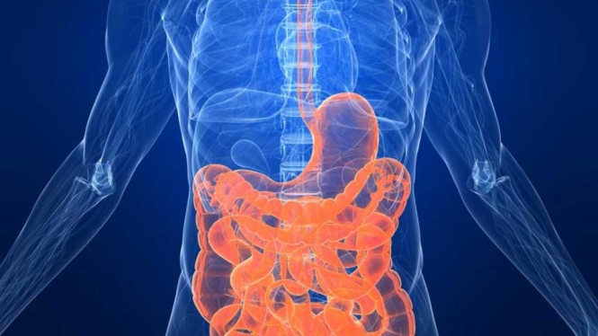
Stomach - the most comprehensive part of the gastrointestinal tract - lies in the left upper abdominal quadrant (stomach area is located below the left costal arch). Its shape is very variable and depends mainly on body position. In general, we distinguish the following sections - cardia (connected with the esophagus), the body of the stomach (middle part) and pylorus part (transition into the duodenum). Domed baggy stomach being its upper pole is called Fundus. In the description of this organ we can not miss its curves: the larger on the left and lower on the right side. The stomach does not only transport the food, but also consumes it. Such a mixed up and "predigested" mush goes into the duodenum, the first section of the small intestine.
Small and large intestine
Duodenum is separated from the stomach by pyloric valve. The small intestine is 4-5 meters long. In addition to the duodenum parts of small intestine are ileum (2/5 upper) and jejunum (3/5 bottom). In the small intestine there occur digestion of food, the further transport and absorption of nutrients. For such a role the folds of the mucosa and circular fibrils are adapted to. The small intestine is separated from the large with ileocaecal valve, which does not pass useless remnants of food from the back. In the large intestine by pumping water and mucus secretion, stool is formed. The large intestine is divided into the cecum and the appendix, colon and rectum with the anal canal. Colon, is divided into ascending, transverse, descending and sigmoid.
Digestive glands: liver and pancreas
During the digestion of food enzymes produced and secreted by the so-called digestive glands are necessary. The best known is probably the liver, located in the right hypochondrium and a large abdomen area. This large organ, weighing from 1300 to 1700 grams, consists of two lobes: right and left. Lobes are divided into lobules. Into the so-called cavity goes blood from the hepatic artery and portal vein (the latter collects blood from the stomach, intestines, spleen and pancreas). The blood leaves the liver, heading to the heart through the hepatic vein. One of the liver functions is the secretion of bile. It collects in the hepatic ducts: right and left, which in turn connect to a recess in the common hepatic duct. The bile does not flow directly into the duodenum, but accumulates in the gallbladder. Cystic duct connects the gallbladder with the common hepatic duct. From that point, the main road leading the bile is called the common bile duct in the nipple larger bills duodenum. Another digestive gland is the pancreas. Enzymes secreted through it are essential for proper digestion of food. The pancreas is composed of the head (covered by a loop of the duodenum), body and tail. Is mainly located on the left side (epigastrium and left hypochondrium). Secretion (juice) of the pancreas is collected (similar to bile in the liver) in the pipes. There are two main lines: pancreatic duct (considered together with the common bile duct in the duodenum) and additional pancreatic duct (considered the lesser duodenal papilla). Just as the liver, pancreatic parenchyma is divided into smaller units - the buds, which secrete digestive enzymes. Between them there are scattered pancreatic islets, which, instead of the duodenum secrete their contents (hormones) into the blood. So they are endocrine gland.
Activity of the digestive system
The role of the digestive system is mainly digestion and absorption of food. These essential functions are connected with others: chyme transport (gastrointestinal motility is responsible for that), the elimination of undigested remains and digestive juices secretion.
Gastrointestinal motility
Motility begins during chewing of food. Contrary to appearances, this is a complicated task divided into six phases. Not everyone seems well aware that the pressure of the jaws (in molar) reaches 150 kg. With the reflex control of the nervous system the soft parts of teeth does not damage. Chewing provides a fragmentation of the food and mixing it with saliva, in which there are already digesting enzymes (starch). Swallowing is equally complicated. In its course, there are three phases: oral, pharyngeal and esophageal. Only the first of them, when food is still in your mouth, you can freely control. Next are reflexive, taking place without the participation of our will. With the coordinated work of many muscles quick and efficient transfer of food bites and fluids to the stomach in such a manner that does not move into the respiratory tract (nose, larynx), or withdrawn back into the esophagus (due to the so-called lower sphincter esophagus) is possible. Muscular layer of the gastrointestinal tract is composed primarily of smooth muscles. This type of tissue is characterized by periodic, rhythmic contractions, which are the basis for all movements leading to the mixing and movement of gastric contents (peristalsis). In various parts of the digestive system we find a different frequency of these movements: in the stomach is about 3/min, and in ileum 8/min. This leads to food content move and then undigested remains up to the rectum, where defecation occurs. In childhood we learn to control this activity and we can suppress it with our will.
Digestion of carbohydrates
The enzymatic digestion of carbohydrates is initiated in the mouth. When food is chewed, crumbled and mixed with saliva produced in the salivary glands containing an enzyme called ptyalin which breaks down some of the bindings in complex carbohydrates. By its action, amylose decomposes into maltose and maltotriose and amylopectin to maltose, maltotriose, and compounds called dextrines built of several molecules of glucose. Ptyalin may act only in the oral cavity and esophagus, as its activity is inhibited in the acidic environment of the stomach. In addition to the ptyalin salivary glands secrete a second enzyme - amylase, which has the ability to hydrolyze complex carbohydrates. It also doesn?t work in an acidic environment. The process of carbohydrates digestion is a multistep process which only begins in the oral cavity. The next steps occur in the subsequent sections of the gastrointestinal tract ? intestines, under the influence of ?-amylase - an enzyme secreted by the pancreas. When the food passes from the stomach into the duodenum, cholecystokinin hormone is secreted by cells lining the walls, causing rich in digestive enzymes pancreatic secretion by the pancreas. It also contains the ?-amylase. It is an enzyme that further breaks down the carbohydrates into quite simple forms, called oligosaccharides. These are long-chain compounds, unbranched, which can be very little absorbed into the blood. Mostly must undergo next step of hydrolysis to sugars, which can now be directly used as an energy source. This process occurs on the surface of cells covering the small intestine with the participation of enzymes called oligosaccharidases and disaccharidases and it is the last stage of this process. We distinguish several types of oligosaccharidases: lactase, maltase, saccharase and izomaltase. Lactase is the only enzyme that allows the hydrolysis of lactose into simple sugars, glucose and galactose. It is already present in fetal life and its activity gradually decreases with age. Lactose is a disaccharide present in large amounts in dairy products. After ingestion is easily degradable in the intestine to simple sugars, glucose and galactose. Lack of the enzyme revealed intolerance of cow's milk and it occurs quite frequently in adults, especially blacks (up to 70% of individuals). Undigested lactose in the colon with the participation of bacteria is converted to hydrogen, methane and carbon dioxide, resulting in persistent bloating, abdominal pain, diarrhea and nausea occurring after the consumption of dairy foods. In this case, the use of restricted diet and the use of milk available in pharmacies lactase preparation greatly reduces or eliminates the symptoms described in their entirety. Maltase is an enzyme that causes the cleavage of glucose from short polymers of glucose. As in the case of lactose found already in utero, and its level remained more or less at the same level throughout life. Saccharase is an enzyme that causes breakdown of sucrose into simple sugars. The amount of it largely depends on the diet. Starvation contributes to a significant decrease in its activity, whereas the carbohydrate rich diet leads to an increase in the amount of this enzyme. This state is also observed in diabetic patients. In this case, the use of drugs that block the activity of sucrose contributes significantly to the reduction of the absorption arising as a result of the action of glucose.
Digestion of proteins
Proteins, in contrast to carbohydrates are much more complex matter of food. They are composed of amino acids linked together by chemical bindings. A large variety of amino acids and related connectivity between them are specific to each organism, both animal and vegetable causes a great diversity of this group. As in case of carbohydrates, those can not be used by the body in the form of the complex - previously must be distributed to individual amino acids. However, this process is only initiated in the stomach. Inside the stomach so called parietal cells are stimulated by nutrient hormones (gastrin, cholecystokinin) and by the nervous system to secrete hydrochloric acid. Its task is twofold. First, it is killing most of microorganisms entering along with the food. Secondly, acting on the protein causes its denaturation, that is destroying chemical bindings in the protein molecule, which determine its complex spatial structure. You can easily tell that the protein is straighten, which facilitates the action of digestive enzymes. In addition to gastric acid, by the so-called main cells of the stomach, enzyme called pepsin is also secreted. Its operation is also possible under acidic conditions and is based on breaking internal bindings of protein, in particular those between amino acids with branched molecules such as leucine and aromatic e.g. tyrosine and phenylalanine. The result of this action is a mixture of different length peptides (peptides are small length chains composed of amino acids) and small amounts of free amino acids. Then the content of predigested food enters the duodenum and so begins the intestinal phase of proteins digestion, which is more complex. Generally it can be divided into three steps: decomposition of protein in the intestine, the distribution of proteins on the surface of cells lining the intestine, which is within the so-called brush border and stage of intracellular digestion. Digestion of proteins in the lumen of the intestine occurs with the participation of enzymes secreted by the pancreas - proteases (trypsin, chymotrypsin and elastase) and pancreatic peptidases (carboxypeptidase A and B). These are the major proteins forming part of the pancreatic juice. Proteases have the ability to break the internal bindings of peptide chains, each of these enzymes have the ability to break down the bonds between specific amino acids: trypsin breaks the binding between the lysine and arginine, chymotrypsin bindings between aromatic amino acids and elastase between amino acids, called aliphatic - valine, glycine, alanine, leucine and isoleucine. Peptidases have the ability to shut off the last amino acid in the peptide chain. In summary, proteases break down to an even shorter peptide chains of amino acids that are then broken down into individual amino acids by peptidases. As a result of these enzymes a mixture of single amino acids and very short chains of amino acids is produced. Now comes the next stage of proteins digestion in so-called brush border. It is a place on the surface of cells lining the intestinal lumen and composed of very many tabs forming a kind of "brush", hence the name. Brush border is a place very rich in exopeptidases ? enzymes decomposing terminal peptide binding in the chain, decomposing dipeptidases second from the end of the peptide bond and bond-degrading endopeptidases in the inner part of the peptide. Digestion in the brush border is the last stage of this process occurring in the intestinal lumen. Its result is a mixture of single amino acids and very short peptides composed of 2-6 amino acids, that are absorbed into cells. Absorption of this is done during a process of using specific active transport systems, which means that the energy consumed during conduct of it. Inside the cell further degradation of short peptides to single amino acids occurs, with the participation of the enzymes contained in cells. When all the peptides are already distributed to individual amino acids, their passive diffusion follows (already without energy input from the body) into the bloodstream, specifically to the portal vein system, which enters the liver. In the liver, there are very complex processes of transformation of amino acids and their further degradation. As you can see, the process of digestion of proteins is very complex and multi-step, and the lack of any enzyme in the course leads to impaired digestion. It can be caused by damage of the organ or cells producing the enzyme and defined as a secondary disorder that is usually found. The second type is known as the primary disorder which is a genetic defect or disturbing the synthesis of several enzymes. An example of secondary disease is celiac disease, which damages the brush border, which significantly reduces the amount of enzymes that are there, which significantly impairs the digestive process. Also several primary disease that interfere with the synthesis of trypsin and brush border enzymes were described.
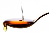
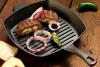
Digestion of fats
Digestion of fats begins in the mouth and stomach, with the participation of linguistic and stomach lipase enzymes secreted by the glands of the base of the tongue, called Ebner glands and stomach glands. But this process is marginal. A significant phase begins in the duodenum. In the stomach, the process prepares fats for digestion, it is about mixing of the slurry and dispersion of ingested fat into small droplets - this phase is called emulsification and occurs through rhythmic stomach contractions called peristaltic movements. After entering the duodenum food gruel is mixed with secreted into light of it bile, which has very strong ability to reduce surface tension, like the washing-up liquid for easy washing greasy dishes. Thus the process of emulsification which began in the stomach is continued, and the fat forms an emulsion consisting of very small droplets of fat. Then, to the lumen of the duodenum separate enzymes produced in the pancreas are getting: pancreatic lipase, phospholipase and esterase. Their task is to hydrolysis, the breakdown of fats complex chemical bindings, which final stage is a mixture of fatty acids. Only in that form fat may be absorbed from the intestine and used by the body as energy or backup material. In diseases of organs producing bile and fat digestive enzymes, often appear so called steatorrhoea. These are mixed with diarrhea, visible to the naked eye drops of fat. This is usually in case of the diseases of pancreas (acute and chronic inflammation), liver and biliary tract.